DNM1L
Protein-coding gene in humans
| DNM1L | |||||||||||||||||||||||||||||||||||||||||||||||||||
|---|---|---|---|---|---|---|---|---|---|---|---|---|---|---|---|---|---|---|---|---|---|---|---|---|---|---|---|---|---|---|---|---|---|---|---|---|---|---|---|---|---|---|---|---|---|---|---|---|---|---|---|
| |||||||||||||||||||||||||||||||||||||||||||||||||||
| Identifiers | |||||||||||||||||||||||||||||||||||||||||||||||||||
| Aliases | DNM1L, DLP1, DRP1, DVLP, DYMPLE, EMPF, HDYNIV, dynamin 1-like, dynamin 1 like, EMPF1, OPA5 | ||||||||||||||||||||||||||||||||||||||||||||||||||
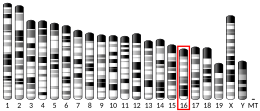
| External IDs | OMIM: 603850; MGI: 1921256; HomoloGene: 6384; GeneCards: DNM1L; OMA:DNM1L - orthologs | ||||||||||||||||||||||||||||||||||||||||||||||||||
| |||||||||||||||||||||||||||||||||||||||||||||||||||
| |||||||||||||||||||||||||||||||||||||||||||||||||||
| |||||||||||||||||||||||||||||||||||||||||||||||||||
| |||||||||||||||||||||||||||||||||||||||||||||||||||
| |||||||||||||||||||||||||||||||||||||||||||||||||||
| Wikidata | |||||||||||||||||||||||||||||||||||||||||||||||||||
| |||||||||||||||||||||||||||||||||||||||||||||||||||


Dynamin-1-like protein is a GTPase that regulates mitochondrial fission. In humans, dynamin-1-like protein, which is typically referred to as dynamin-related protein 1 (Drp1), is encoded by the DNM1L gene and is part of the dynamin superfamily (DSP) family of proteins.[5][6][7]
Structure
Drp1, which is a member of the dynamin superfamily of proteins, consists of a GTPase and GTPase effector domain that are separated from each other by a helical segment of amino acids.[8] There are 3 mouse and 6 human isoforms of Drp1, including a brain-specific variant.[9] Drp1 exists as homooligomers[10] and its function relies on its oligomerization ability.[11]
Function
Mitochondria routinely undergo fission and fusion events that maintain a dynamic reticular network. Drp1 is a fundamental component of mitochondrial fission.[12] Indeed, Drp1 deficient neurons have large, strongly interconnected mitochondria[13] due to dysfunctional fission machinery. Fission helps facilitate mitophagy, which is the breakdown and recycling of damaged mitochondria. Dysfunction in the DRP activity may result in mutated DNA or malfunctioning proteins diffusing throughout the mitochondrial system. In addition, fission results in fragmented mitochondria more capable of producing of reactive oxygen species, which can disrupt normal biochemical processes inside of cells.[14] ROS can be formed from incomplete transfer of electrons through the electron transport chain. Furthermore, fission influences calcium flux within the cell, linking Drp1 to apoptosis and cancer.[15]
Several studies have indicated that Drp1 is essential for proper embryonic development. Drp1 knockout mice exhibit abnormal brain development and die around embryonic day 12. In neural specific Drp1 knockout mice, brain size is reduced and apoptosis is increased. Synapse formation and neurite growth are also impaired. A second group of researchers generated another neural specific knockout mouse line. They found that knocking out Drp1 resulted in the appearance of large mitochondria in Purkinje cells and prevented neural tube formation.[9]
In humans, loss of Drp1 function affects brain development and is also associated with early mortality.[8]
Interactions
The majority of knowledge about mitochondrial fission comes from studies with yeast. The yeast homolog of Drp1 is dynamin-1 (Dnm1), which interacts with Fis1 through Mdv1. This interaction causes Dnm1 to oligomerize and form rings around dividing mitochondria at the so-called "constriction point".[8][16] Drp1 has also been shown to interact with GSK3B.[6] In mammals, Drp1 receptors include Mff, Mid49 and Mid51[17][18]
Post-translational modifications to Drp1 (e.g. phosphorylation) can alter its activity and affect the rate of fission.[19]
Drp1 has two major phosphorylation sites. The CDK phosphorylation site is S579, and the PKA site is S600 in Drp1 isoform 3. Phosphorylation by CDK is thought to be activating, whereas PKA phosphorylation is thought to be inhibitory. Recently, CaMKII was shown to phosphorylate Drp1 at S616. This was shown to occur in response to chronic Beta-adrenergic stimulation and to promote mPTP opening.[20] Other post-translational modifications include S-nitrosylation, sumoylation, and ubiquitination. Higher S- nitrosylation modifications of Drp1, which enhances Drp1 activity, have been observed in Alzheimer’s Disease. Furthermore, Drp1 has been shown to interact with Aβ monomers, thought to play an important role in Alzheimer’s Disease, exacerbating the disease and its symptoms.[21] Drp1 has been linked to a number of pathways and processes including cell division, apoptosis, and necrosis. Drp1 has been shown to stabilize p53 during oxidative stress, promoting its translocation to the mitochondria and encouraging mitochondrial- related necrosis.[22] In addition, cyclin B1- CDK activates Drp1, causing fragmentation and ensuring mitochondria are distributed to each daughter cell after mitosis. Likewise, different transcriptional controllers are able to alter Drp1 activity through gene expression and regulation. For example, PPARGC1A and [HIF1A] regulated Drp1 activity through gene expression.[14]
Therapy
Inhibition of Drp1 has been considered for possible therapeutics for a variety of diseases. The most studied inhibitor is a small molecule named mitochondrial division inhibitor 1 (mdivi-1) which may have off-target effects such as inhibition of complex 1 of the mitochondrial respiratory chain.[23] The inhibitors putative function is preventing the GTPase activity of Drp1 thus preventing the activation and localization to the mitochondria.[14] Midiv-1 has been demonstrated to attenuate the effects of ischemia reperfusion injury after cardiac arrest. The treatment prevented both mitochondria fragmentation and increased cell viability.[24] Similarly, midiv-1 has demonstrated neuroprotective effects by greatly reducing neuron death due to seizure. Furthermore, the study showed midiv-1 was capable to preventing the activation of caspase 3 by reversing the release of cytochrome c in intrinsic apoptosis.[25] Whether mdivi-1 inhibits Drp1 or not, its therapeutic potential is certainly evident. Other than directly inhibiting Drp1, certain inhibitors of proteins involved in the posttranslational modifications of Drp1 have been studied. FK506 is a calcineurin inhibitor, which functions to dephosphorylate the serine 637 position of Drp1, encouraging translocation to the mitochondria and fragmentation. FK506 was shown to also preserve mitochondrial morphology after reperfusion injury.[24]
References
- ^ a b c GRCh38: Ensembl release 89: ENSG00000087470 – Ensembl, May 2017
- ^ a b c GRCm38: Ensembl release 89: ENSMUSG00000022789 – Ensembl, May 2017
- ^ "Human PubMed Reference:". National Center for Biotechnology Information, U.S. National Library of Medicine.
- ^ "Mouse PubMed Reference:". National Center for Biotechnology Information, U.S. National Library of Medicine.
- ^ Shin HW, Shinotsuka C, Torii S, Murakami K, Nakayama K (September 1997). "Identification and subcellular localization of a novel mammalian dynamin-related protein homologous to yeast Vps1p and Dnm1p". Journal of Biochemistry. 122 (3): 525–30. doi:10.1093/oxfordjournals.jbchem.a021784. PMID 9348079.
- ^ a b Hong YR, Chen CH, Cheng DS, Howng SL, Chow CC (August 1998). "Human dynamin-like protein interacts with the glycogen synthase kinase 3beta". Biochemical and Biophysical Research Communications. 249 (3): 697–703. doi:10.1006/bbrc.1998.9253. PMID 9731200.
- ^ "Entrez Gene: DNM1L dynamin 1-like".
- ^ a b c Westermann B (December 2010). "Mitochondrial fusion and fission in cell life and death". Nature Reviews. Molecular Cell Biology. 11 (12): 872–84. doi:10.1038/nrm3013. PMID 21102612. S2CID 3342603.
- ^ a b Reddy PH, Reddy TP, Manczak M, Calkins MJ, Shirendeb U, Mao P (June 2011). "Dynamin-related protein 1 and mitochondrial fragmentation in neurodegenerative diseases". Brain Research Reviews. 67 (1–2): 103–18. doi:10.1016/j.brainresrev.2010.11.004. PMC 3061980. PMID 21145355.
- ^ Kwapiszewska K, Kalwarczyk T, Michalska B, Szczepański K, Szymański J, Patalas-Krawczyk P, Andryszewski T, Iwan M, Duszyński J, Hołyst R (April 2019). "Determination of oligomerization state of Drp1 protein in living cells at nanomolar concentrations". Scientific Reports. 9 (1): 5906. Bibcode:2019NatSR...9.5906K. doi:10.1038/s41598-019-42418-0. PMC 6459820. PMID 30976093.
- ^ Michalska BM, Kwapiszewska K, Szczepanowska J, Kalwarczyk T, Patalas-Krawczyk P, Szczepański K, Hołyst R, Duszyński J, Szymański J (May 2018). "Insight into the fission mechanism by quantitative characterization of Drp1 protein distribution in the living cell". Scientific Reports. 8 (1): 8122. Bibcode:2018NatSR...8.8122M. doi:10.1038/s41598-018-26578-z. PMC 5970238. PMID 29802333.
- ^ Smirnova E, Shurland DL, Ryazantsev SN, van der Bliek AM (October 1998). "A human dynamin-related protein controls the distribution of mitochondria". The Journal of Cell Biology. 143 (2): 351–8. doi:10.1083/jcb.143.2.351. PMC 2132828. PMID 9786947.
- ^ Wiemerslage L, Lee D (March 2016). "Quantification of mitochondrial morphology in neurites of dopaminergic neurons using multiple parameters". Journal of Neuroscience Methods. 262: 56–65. doi:10.1016/j.jneumeth.2016.01.008. PMC 4775301. PMID 26777473.
- ^ a b c Archer SL (December 2013). "Mitochondrial dynamics--mitochondrial fission and fusion in human diseases". The New England Journal of Medicine. 369 (23): 2236–51. doi:10.1056/NEJMra1215233. PMID 24304053. S2CID 2346449.
- ^ Zhang C, Yuan XR, Li HY, Zhao ZJ, Liao YW, Wang XY, Su J, Sang SS, Liu Q (January 2014). "Downregualtion of dynamin-related protein 1 attenuates glutamate-induced excitotoxicity via regulating mitochondrial function in a calcium dependent manner in HT22 cells". Biochemical and Biophysical Research Communications. 443 (1): 138–43. doi:10.1016/j.bbrc.2013.11.072. PMID 24284040.
- ^ Lackner LL, Horner JS, Nunnari J (August 2009). "Mechanistic analysis of a dynamin effector". Science. 325 (5942): 874–7. Bibcode:2009Sci...325..874L. doi:10.1126/science.1176921. PMC 6546417. PMID 19679814.
- ^ Otera H, Wang C, Cleland MM, Setoguchi K, Yokota S, Youle RJ, Mihara K (December 2010). "Mff is an essential factor for mitochondrial recruitment of Drp1 during mitochondrial fission in mammalian cells". The Journal of Cell Biology. 191 (6): 1141–58. doi:10.1083/jcb.201007152. PMC 3002033. PMID 21149567.
- ^ Palmer CS, Osellame LD, Laine D, Koutsopoulos OS, Frazier AE, Ryan MT (June 2011). "MiD49 and MiD51, new components of the mitochondrial fission machinery". EMBO Reports. 12 (6): 565–73. doi:10.1038/embor.2011.54. PMC 3128275. PMID 21508961.
- ^ Knott AB, Perkins G, Schwarzenbacher R, Bossy-Wetzel E (July 2008). "Mitochondrial fragmentation in neurodegeneration". Nature Reviews. Neuroscience. 9 (7): 505–18. doi:10.1038/nrn2417. PMC 2711514. PMID 18568013.
- ^ Xu S, Wang P, Zhang H, Gong G, Gutierrez Cortes N, Zhu W, Yoon Y, Tian R, Wang W (October 2016). "CaMKII induces permeability transition through Drp1 phosphorylation during chronic β-AR stimulation". Nature Communications. 7: 13189. Bibcode:2016NatCo...713189X. doi:10.1038/ncomms13189. PMC 5067512. PMID 27739424.
- ^ Yan MH, Wang X, Zhu X (September 2013). "Mitochondrial defects and oxidative stress in Alzheimer disease and Parkinson disease". Free Radical Biology & Medicine. 62: 90–101. doi:10.1016/j.freeradbiomed.2012.11.014. PMC 3744189. PMID 23200807.
- ^ Guo X, Sesaki H, Qi X (July 2014). "Drp1 stabilizes p53 on the mitochondria to trigger necrosis under oxidative stress conditions in vitro and in vivo". The Biochemical Journal. 461 (1): 137–46. doi:10.1042/BJ20131438. PMC 4381936. PMID 24758576.
- ^ Bordt EA, Clerc P, Roelofs BA, Saladino AJ, Tretter L, Adam-Vizi V, Cherok E, Khalil A, Yadava N, Ge SX, Francis TC, Kennedy NW, Picton LK, Kumar T, Uppuluri S, Miller AM, Itoh K, Karbowski M, Sesaki H, Hill RB, Polster BM (March 2017). "The Putative Drp1 Inhibitor mdivi-1 Is a Reversible Mitochondrial Complex I Inhibitor that Modulates Reactive Oxygen Species". Developmental Cell. 40 (6): 583–594.e6. doi:10.1016/j.devcel.2017.02.020. PMC 5398851. PMID 28350990.
- ^ a b Sharp WW, Fang YH, Han M, Zhang HJ, Hong Z, Banathy A, Morrow E, Ryan JJ, Archer SL (January 2014). "Dynamin-related protein 1 (Drp1)-mediated diastolic dysfunction in myocardial ischemia-reperfusion injury: therapeutic benefits of Drp1 inhibition to reduce mitochondrial fission". FASEB Journal. 28 (1): 316–26. doi:10.1096/fj.12-226225. PMC 3868827. PMID 24076965.
- ^ Xie N, Wang C, Lian Y, Zhang H, Wu C, Zhang Q (June 2013). "A selective inhibitor of Drp1, mdivi-1, protects against cell death of hippocampal neurons in pilocarpine-induced seizures in rats". Neuroscience Letters. 545: 64–8. doi:10.1016/j.neulet.2013.04.026. PMID 23628672. S2CID 46558819.
Further reading
- Pawlikowska P, Orzechowski A (2007). "[Role of transmembrane GTPases in mitochondrial morphology and activity]". Postepy Biochemii. 53 (1): 53–9. PMID 17718388.
- Kamimoto T, Nagai Y, Onogi H, Muro Y, Wakabayashi T, Hagiwara M (January 1998). "Dymple, a novel dynamin-like high molecular weight GTPase lacking a proline-rich carboxyl-terminal domain in mammalian cells". The Journal of Biological Chemistry. 273 (2): 1044–51. doi:10.1074/jbc.273.2.1044. PMID 9422767.
- Imoto M, Tachibana I, Urrutia R (May 1998). "Identification and functional characterization of a novel human protein highly related to the yeast dynamin-like GTPase Vps1p". Journal of Cell Science. 111 ( Pt 10) (10): 1341–9. doi:10.1242/jcs.111.10.1341. PMID 9570752.
- Rasmussen RK, Rusak J, Price G, Robinson PJ, Simpson RJ, Dorow DS (October 1998). "Mixed-lineage kinase 2-SH3 domain binds dynamin and greatly enhances activation of GTPase by phospholipid". The Biochemical Journal. 335 ( Pt 1) (Pt 1): 119–24. doi:10.1042/bj3350119. PMC 1219759. PMID 9742220.
- Smirnova E, Shurland DL, Ryazantsev SN, van der Bliek AM (October 1998). "A human dynamin-related protein controls the distribution of mitochondria". The Journal of Cell Biology. 143 (2): 351–8. doi:10.1083/jcb.143.2.351. PMC 2132828. PMID 9786947.
- Smirnova E, Griparic L, Shurland DL, van der Bliek AM (August 2001). "Dynamin-related protein Drp1 is required for mitochondrial division in mammalian cells". Molecular Biology of the Cell. 12 (8): 2245–56. doi:10.1091/mbc.12.8.2245. PMC 58592. PMID 11514614.
- Karbowski M, Lee YJ, Gaume B, Jeong SY, Frank S, Nechushtan A, Santel A, Fuller M, Smith CL, Youle RJ (December 2002). "Spatial and temporal association of Bax with mitochondrial fission sites, Drp1, and Mfn2 during apoptosis". The Journal of Cell Biology. 159 (6): 931–8. doi:10.1083/jcb.200209124. PMC 2173996. PMID 12499352.
- Koch A, Thiemann M, Grabenbauer M, Yoon Y, McNiven MA, Schrader M (March 2003). "Dynamin-like protein 1 is involved in peroxisomal fission". The Journal of Biological Chemistry. 278 (10): 8597–605. doi:10.1074/jbc.M211761200. PMID 12499366.
- Li X, Gould SJ (May 2003). "The dynamin-like GTPase DLP1 is essential for peroxisome division and is recruited to peroxisomes in part by PEX11". The Journal of Biological Chemistry. 278 (19): 17012–20. doi:10.1074/jbc.M212031200. PMID 12618434.
- Breckenridge DG, Stojanovic M, Marcellus RC, Shore GC (March 2003). "Caspase cleavage product of BAP31 induces mitochondrial fission through endoplasmic reticulum calcium signals, enhancing cytochrome c release to the cytosol". The Journal of Cell Biology. 160 (7): 1115–27. doi:10.1083/jcb.200212059. PMC 2172754. PMID 12668660.
- Yoon Y, Krueger EW, Oswald BJ, McNiven MA (August 2003). "The mitochondrial protein hFis1 regulates mitochondrial fission in mammalian cells through an interaction with the dynamin-like protein DLP1". Molecular and Cellular Biology. 23 (15): 5409–20. doi:10.1128/MCB.23.15.5409-5420.2003. PMC 165727. PMID 12861026.
- Howng SL, Sy WD, Cheng TS, Lieu AS, Wang C, Tzou WS, Cho CL, Hong YR (February 2004). "Genomic organization, alternative splicing, and promoter analysis of human dynamin-like protein gene". Biochemical and Biophysical Research Communications. 314 (3): 766–72. doi:10.1016/j.bbrc.2003.12.172. PMID 14741701.
- Lee YJ, Jeong SY, Karbowski M, Smith CL, Youle RJ (November 2004). "Roles of the mammalian mitochondrial fission and fusion mediators Fis1, Drp1, and Opa1 in apoptosis". Molecular Biology of the Cell. 15 (11): 5001–11. doi:10.1091/mbc.E04-04-0294. PMC 524759. PMID 15356267.
- Pitts KR, McNiven MA, Yoon Y (November 2004). "Mitochondria-specific function of the dynamin family protein DLP1 is mediated by its C-terminal domains". The Journal of Biological Chemistry. 279 (48): 50286–94. doi:10.1074/jbc.M405531200. PMID 15364948.
- Goehler H, Lalowski M, Stelzl U, Waelter S, Stroedicke M, Worm U, Droege A, Lindenberg KS, Knoblich M, Haenig C, Herbst M, Suopanki J, Scherzinger E, Abraham C, Bauer B, Hasenbank R, Fritzsche A, Ludewig AH, Büssow K, Buessow K, Coleman SH, Gutekunst CA, Landwehrmeyer BG, Lehrach H, Wanker EE (September 2004). "A protein interaction network links GIT1, an enhancer of huntingtin aggregation, to Huntington's disease". Molecular Cell. 15 (6): 853–65. doi:10.1016/j.molcel.2004.09.016. PMID 15383276.
- Germain M, Mathai JP, McBride HM, Shore GC (April 2005). "Endoplasmic reticulum BIK initiates DRP1-regulated remodelling of mitochondrial cristae during apoptosis". The EMBO Journal. 24 (8): 1546–56. doi:10.1038/sj.emboj.7600592. PMC 1142564. PMID 15791210.
- Narayanan R, Leonard M, Song BD, Schmid SL, Ramaswami M (April 2005). "An internal GAP domain negatively regulates presynaptic dynamin in vivo: a two-step model for dynamin function". The Journal of Cell Biology. 169 (1): 117–26. doi:10.1083/jcb.200502042. PMC 2171915. PMID 15824135.
External links
- DNM1L human gene location in the UCSC Genome Browser.
- DNM1L human gene details in the UCSC Genome Browser.
- Overview of all the structural information available in the PDB for UniProt: O00429 (Dynamin-1-like protein) at the PDBe-KB.


